
Not every unusual gel result is caused by poor sample quality or experimental mistakes. Sometimes, your agarose gel is simply revealing the hidden physics of gel electrophoresis. One such phenomenon is the appearance of smiling DNA bands—a common artifact that tells an important story about heat, voltage, and DNA migration.
Why Do DNA Bands Smile? The Hidden Physics Behind Curved DNA Bands in Agarose Gel Electrophoresis
“Your DNA is smiling—it is trying to tell you something.”
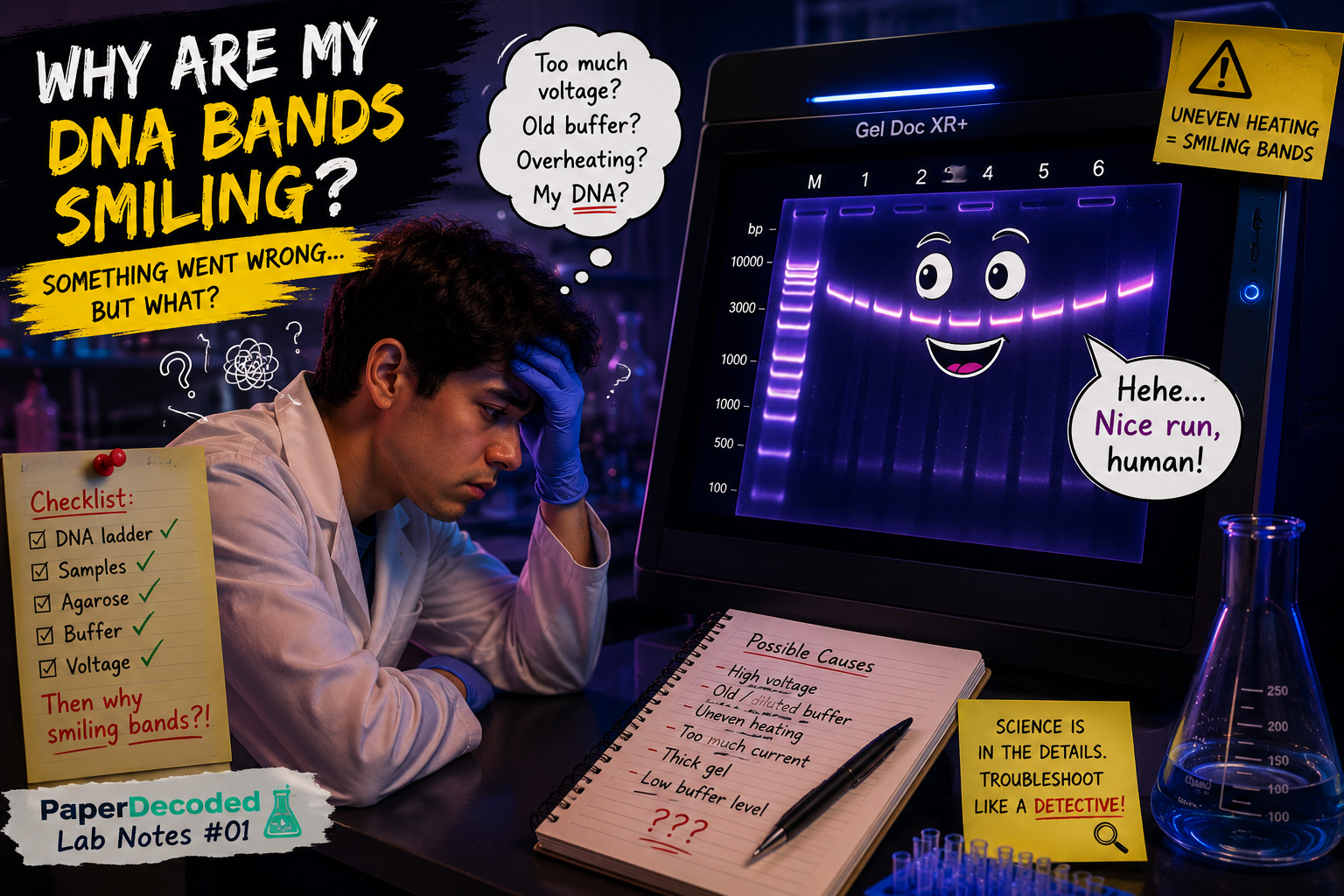
You Did Everything Right… So Why Are Your DNA Bands Smiling?
You prepared fresh agarose gel after a molecular biology assay like PCR. You added the correct amount of loading dye. The DNA ladder looked perfectly normal. You connected the electrodes correctly and ran the gel at 120 V. But when you finally placed the gel under the UV transilluminator, something looked strange. Instead of forming straight horizontal bands, every lane curved upward like a smile. If you’ve ever seen this, don’t worry—you are not alone. Smiling DNA bands are one of the most common electrophoresis artifacts in molecular biology laboratories. More importantly, they are not random. Your gel is revealing a hidden story about electricity, heat, and the physics of electrophoresis. Today, let’s decode that story.
What Does a “Smiling Band” Look Like?
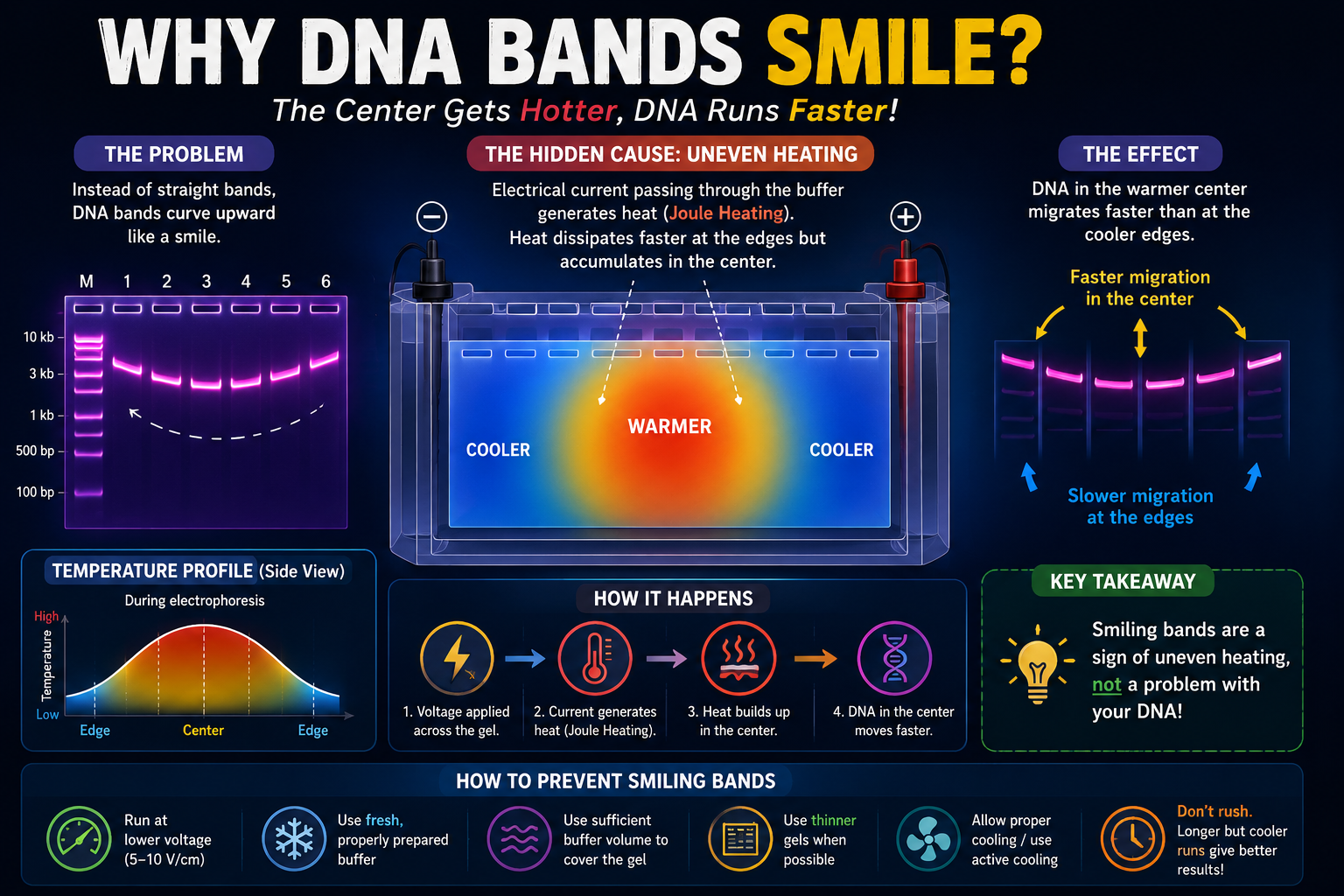
Instead of appearing as a straight horizontal line, the DNA band appears curved. The DNA in the center lanes has migrated slightly farther than the DNA near the edges. The result resembles a smile. Interestingly, this distortion may also affects all lanes, including the DNA ladder. That tells us the problem is with the electrophoresis conditions—not with your DNA samples or errors during DNA purification from PCR product or Plasmid DNA isolation or Genomic DNA isolation or cDNA preparation.
First, Remember How DNA Normally Migrates
Under ideal conditions, every DNA fragment experiences the same electric field across the Agarose Gel. Because DNA carries a uniformly negative charge, every fragment migrates toward the positive electrode (anode).
If the electric field remains uniform, identical DNA fragments in different lanes travel exactly the same distance, producing perfectly straight bands.
So why does this suddenly change?
The Hidden Culprit: Heat
Most of the time a researcher may think electrophoresis is simply about applying voltage and check this on Gel-Doc system. In reality, every electrophoresis experiment is also a heating experiment. Whenever electrical current passes through the conductive buffer, some electrical energy is converted into heat. Physicists call this Joule heating. The amount of heat generated depends mainly on:
- Applied voltage
- Electrical current
- Buffer conductivity
- Duration of the run
This heating is unavoidable. The question is whether it remains uniform.
Why Does the Middle of the Gel Become Hotter?
This is the fascinating part. The gel edges are surrounded by buffer and the plastic walls of the electrophoresis chamber, allowing heat to dissipate more efficiently. The center of the gel has fewer pathways to lose heat. As electrophoresis continues, the middle region gradually becomes warmer than the edges.
Even a temperature difference of only a few degrees can influence DNA migration.
Why Does Heat Make DNA Run Faster?
A warmer agarose gel behaves slightly differently. As temperature increases:
- Buffer viscosity decreases.
- DNA experiences less resistance.
- Molecular movement becomes easier.
- DNA migrates slightly faster.
The DNA fragments in the warmer center therefore travel farther than identical fragments near the cooler edges. The result is a characteristic upward curve—a “smile.”
Think of three runners racing on different roads. The runners at the edges are on rough pavement. The runner in the center is on freshly paved asphalt. Everyone runs with the same effort, but the smoother road allows the center runner to finish slightly ahead. Your DNA behaves in much the same way.
Other Reasons Your Bands May Smile
Heat is the most common cause, but it is not the only one.
1. Running the Gel at Excessively High Voltage
Many researchers increase the voltage to finish sooner. Unfortunately, higher voltage dramatically increases Joule heating. The gel runs faster—but band quality often suffers. As a general guideline, electrophoresis is usually performed at 5–10 V per centimeter (distance between electrodes). Exceeding this range increases the risk of overheating, smiling bands, and reduced resolution.
2. High-Ionic-Strength Buffer
Using an incorrectly prepared or overly concentrated TAE/TBE buffer increases conductivity. Higher conductivity means more current. More current means more heat. More heat increases the likelihood of smiling bands in Agarose Gel.
3. Reusing Buffer Too Many Times
Old running buffer may no longer have uniform ionic composition. Electrolysis during previous runs can alter pH and ion distribution, while evaporation can change concentration. These changes make the electric field less uniform and increase the chance of distorted migration. Fresh buffer often restores consistent results.
4. Large or Thick Gels
Large gels retain heat more efficiently than smaller ones. Thicker gels also dissipate heat more slowly. Without sufficient cooling, temperature gradients become more pronounced.
How Can You Prevent Smiling Bands?
Fortunately, the solution is usually simple.
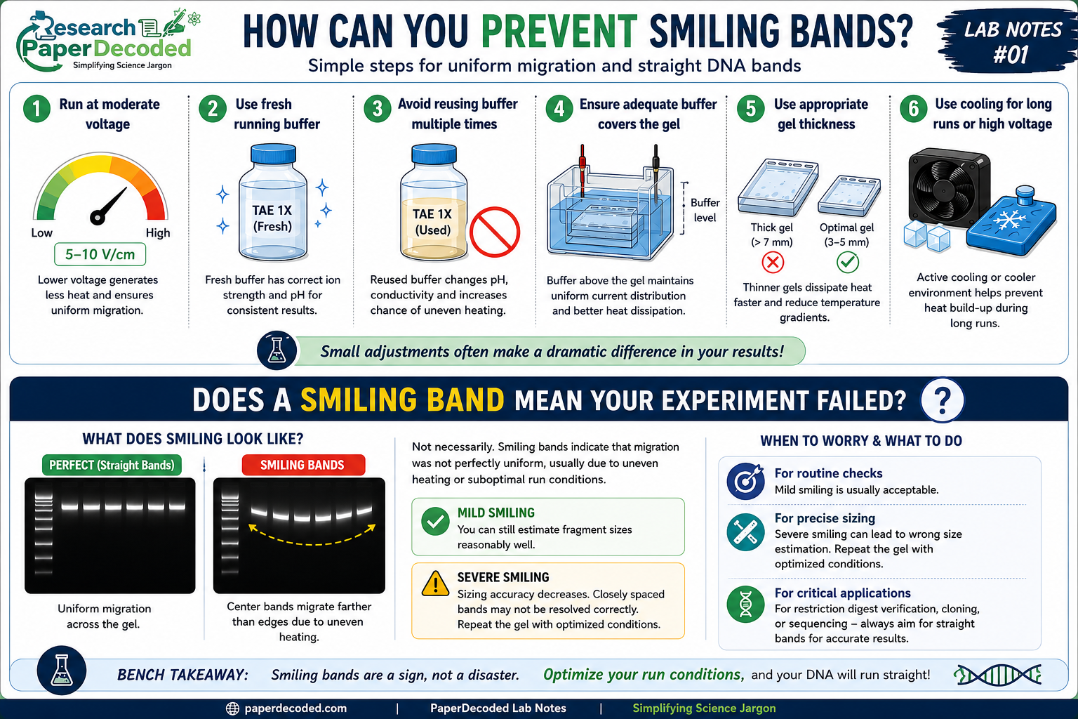
- Run gels at moderate voltage rather than the maximum possible.
- Prepare fresh running buffer with the correct concentration.
- Avoid repeatedly reusing electrophoresis buffer.
- Allow adequate buffer to completely cover the gel.
- Reduce gel thickness when possible.
- For long electrophoresis runs, consider active cooling or performing the run in a cooler environment.
- Small adjustments often make a dramatic difference.
Does a Smiling Band Mean Your Experiment Failed?
Not necessarily. Smiling bands mainly indicate that migration of your DNA samples in agarose gel electrophoresis was not perfectly uniform. If the curvature is mild, you may still be able to determine fragment sizes accurately. However, severe smiling can reduce sizing accuracy and make closely spaced DNA fragments difficult to distinguish. For applications requiring precise fragment analysis—such as restriction digest verification or DNA sizing—it’s best to repeat the gel under optimized conditions.
PaperDecoded Insight
Most people think electrophoresis is simply “DNA moving through a gel.” In reality, it is a balance of electricity, heat transfer, fluid properties, and polymer physics. A smiling DNA band is not merely a cosmetic defect. It is evidence that your gel developed an uneven temperature landscape. Your DNA simply followed the easiest path. The gel was quietly revealing the physics happening beneath the surface.
Bench Takeaway
When DNA bands smile, don’t blame the DNA or yourself. In most cases, the real culprit is uneven heating caused by excessive voltage, incorrect buffer conditions, or poor heat dissipation. Straight bands are often the reward for good electrophoresis physics, not just good molecular biology.
Want to Explore the Science Further?
Key Research Papers
- Zarei, M. et al. (2015) ‘Improvement of heat dissipation in agarose gel electrophoresis by metal oxide nanoparticles,’ RSC Advances, 5(108), pp. 88655–88665. https://doi.org/10.1039/c5ra19678g.
- Serwer, P. (1983) ‘Agarose gels: Properties and use for electrophoresis,’ Electrophoresis, 4(6), pp. 375–382. https://doi.org/10.1002/elps.1150040602.
Sanderson, B.A. et al. (2014) ‘Modification of gel architecture and TBE/TAE buffer composition to minimize heating during agarose gel electrophoresis,’ Analytical Biochemistry, 454, pp. 44–52. https://doi.org/10.1016/j.ab.2014.03.003.

Editor, Research Paper Decoded