
Quick Note:
DNA is negatively charged and the electric field completed with TAE/TBE buffer allow them to move according to their size through EtBr stained agarose gel. The EtBr molecule chelated with DNA gets visible under UV exposer. The smaller sized DNA moves fast and appeared near the end of agarose gel.
The DNA Highway Every Biologist Uses but few Truly Understand
You have probably done agarose gel electrophoresis dozens of times.
Every time after experimental planning any PhD student measure agarose, dissolve in 1X TAE buffer and warm in microwave and cast the gel with EtBr staining. Then samples being loaded according to the experimental design and run at 100 volt for 30 minutes.
For many UG students, it is a practical class ritual. For PG students, it becomes a weekly habit. For PhD scholars and professors, it is often an everyday experiment after a PCR or DNA isolation and other molecular biology related experiments. Yet a surprising question remains:
How many of us actually understand what is happening inside that transparent slab of gel?
A research project nurtured daily with minute troubleshooting. Experimental troubleshooting always ask for conceptual clarity about the experiment performed. Before you fix any issue you must know why agarose is used why not starch or gelatin or something else? Why does the DNA moves? Why they moves according to their size? Why EtBr is used and what does the UV light do so that they appeared under UV transilluminator? And also:
What will be happen if someone loaded proteins on an agarose gel instead of DNA?
Today, let us stop following protocols blindly and understand the hidden science behind one of the most common experiments performed daily in a molecular biology lab.

Imagine DNA Running Through a Forest
Suppose someone asks you to run across an open football field. It’s easy to run. Now imagine running through a dense forest filled with trees. It would be much harder. Now imagine a giant elephant and a rabbit trying to cross that forest. The rabbit can weave through spaces quickly. The elephant struggles.
This simple story explains agarose gel electrophoresis. DNA molecules travel through a mesh-like agarose network. Small DNA fragments move quickly through tiny pores. Larger DNA fragments struggle and move slowly. That is why after electrophoresis, DNA fragments separate according to size. Smaller DNA fragments travel farther. Larger fragments remain behind. The agarose gel essentially acts like a molecular obstacle course.
What Is Agarose Gel Electrophoresis?
Agarose gel electrophoresis is a technique used to separate nucleic acid molecules primarily based on size under the influence of an electric field.
DNA molecules contains negatively charged phosphate groups. When electricity is applied:
- DNA moves toward the positive electrode (anode)
- Agarose creates resistance
- Smaller fragments travel faster
- Larger fragments travel slower
After separation, DNA bands become visible using fluorescent dyes. Simple. But beneath this simplicity lies clever molecular engineering.
Where Does Agarose Come From?
Agarose is not a synthetic laboratory material. Actually, agarose comes from seaweed. It is purified from red algae. When heated in buffer and cooled, agarose molecules organize into a three-dimensional network containing pores. These pores become molecular tunnels for DNA movement. More the agarose concentration less the pore size. Nature unknowingly created a near-perfect material for DNA separation.
Why Agarose? Why Not Any Other Material?
This question is rarely discussed. If all we need is a porous material, why not use gelatin, starch, collagen, or synthetic polymers? The answer lies in several unique properties of agarose.
1. Agarose Forms Uniform Pores: The pore sizes are highly reproducible. Uniform pores mean predictable migration. If pores varied randomly, DNA bands would smear badly.
2. Agarose Is Chemically Inert: DNA should move through the matrix without interacting chemically. Agarose behaves like a silent observer. It simply creates pathways. It does not strongly bind DNA.
3. Easy Gel Preparation: Heat. Cool. Pour. Done. No toxic polymerization reactions. No catalyst. No complicated chemistry. This simplicity made agarose the universal favorite.
4. Adjustable Pore Size: Changing agarose concentration changes pore size. 0.7% agarose: Large pores Good for large DNA fragments. 2–3% agarose: Small pores Good for tiny PCR products. Researchers can tune separation simply by changing agarose concentration. Very few materials offer this flexibility.
Why Does DNA Always Move Toward the Positive Side?
DNA is a negatively charged polymer. Each nucleotide contributes phosphate groups. These phosphates remain negatively charged at physiological pH. Therefore the entire DNA molecule behaves like a negatively charged rope.
Electric fields pull negative molecules toward the positive electrode. That is why DNA consistently migrates in one direction. Many students memorize this fact.
Now think of this interesting factor. Larger DNA contains more phosphate groups. Shouldn’t larger DNA move faster?
Interesting answer: Longer DNA gains:
- more negative charge
- but also more mass
Charge and mass increase proportionally. Therefore agarose pore resistance becomes the major separating factor.
The Buffer is Doing More Work Than You Think
Students usually focus on gel preparation and DNA loading. Buffers often receive little attention. But imagine running electricity without ions. Current would collapse. Buffers like TAE and TBE provide ions that carry electrical current. They also:
- maintain pH
- prevent DNA degradation
- stabilize migration
- reduce overheating
Without buffer, your gel experiment would fail almost instantly. The buffer is the hidden infrastructure of electrophoresis. But what exactly makes this invisible infrastructure work? Let us dive inside the molecular recipe behind agarose electrophoresis.
Think of TAE Buffer (Tris Acetate EDTA) as a tiny molecular city where every component has a different job.
Tris Base: The pH bodyguard. Tris base in TAE buffer maintains the pH around 8. DNA phosphates remain ionized and maintain stable charge behavior under electrophoresis conditions.
Acetate: The current carrier. Acetate provides the ions helping electrical current flow during agarose gel electrophoresis.
EDTA: Molecular security guard. Many nucleases need magnesium. EDTA captures magnesium, which reduces the free magnesium which results in less DNA degradation.
A strange laboratory question now appears: if TAE is simply a buffer, why not borrow protein gel buffer?
Why not use SDS-PAGE running buffer? Protein buffers commonly contain Tris-Glycine-SDS. SDS behaves as a detergent. Proteins need SDS because proteins naturally vary in:
- charge
- shape
- folding
SDS unfolds proteins and coats them with negative charge. DNA already possesses strong negative charge. DNA does not need this treatment. If protein buffer is used:
- conductivity changes
- heating may increase
- migration changes
- SDS may alter interactions
- downstream applications can suffer
Protocols are carefully designed ecosystems. Changing one component creates chain reactions.
Why Use Ethidium Bromide?
Now comes the famous orange chemical that everyone fears.
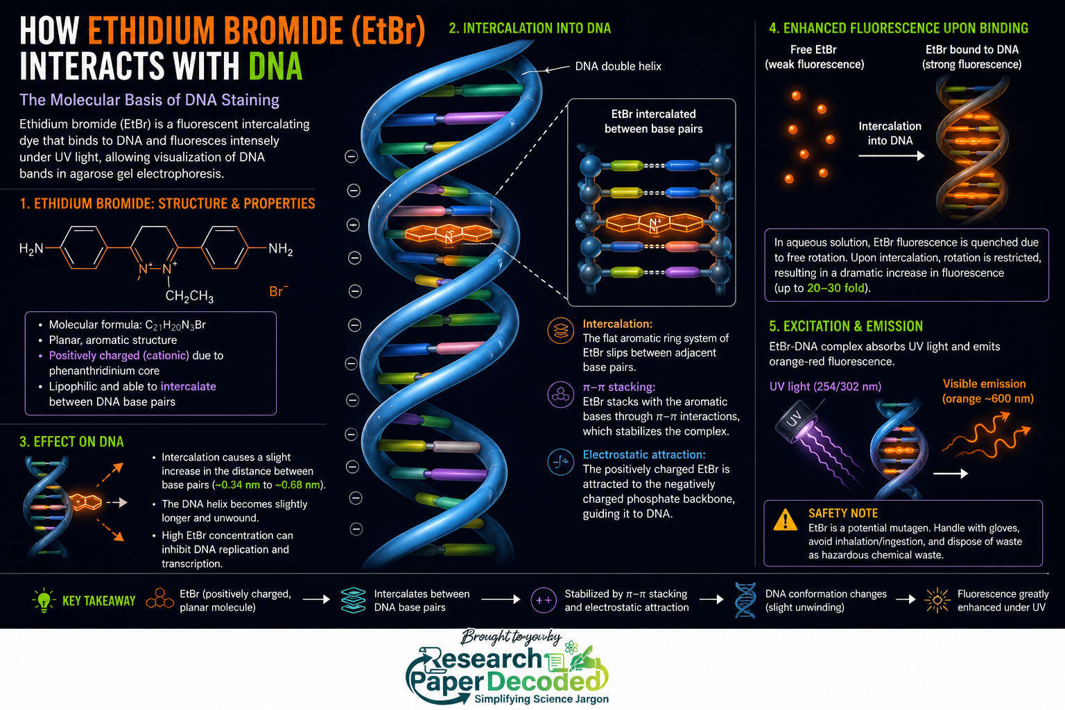
Ethidium bromide. Most students learn: “EtBr stains DNA.” But this sentence hides fascinating molecular behavior. Ethidium bromide is a flat aromatic molecule. DNA contains stacked bases like pages inside a book. EtBr slips between these pages. Once trapped inside DNA, its fluorescence changes dramatically. But why can EtBr enter DNA so easily? The answer hides in geometry. EtBr contains flat aromatic rings. DNA bases are also flat and stacked. Flat molecules often prefer stacking with other flat molecules through π–π interactions. Scientists call this: Intercalation. EtBr also carries a positive charge, which helps attract it toward DNA’s negatively charged phosphate backbone before intercalation stabilizes the interaction.
Free EtBr fluoresces weakly. DNA-bound EtBr glows intensely. That is why DNA suddenly becomes visible. EtBr itself does not create DNA. It simply acts like a molecular highlighter.
Why Does DNA-Bound EtBr Glow More?
This is where physics enters biology. Free EtBr molecules in solution rotate and vibrate freely. This motion wastes absorbed energy. When EtBr inserts into DNA, movement becomes restricted. Now more absorbed energy gets released as fluorescence. Result: Strong orange emission. DNA effectively locks EtBr into a rigid position and turns it into a brighter light source.
Why Use UV Light Specifically?
Let’s come to an interesting question: “Why not shine normal white light?”
EtBr strongly absorbs ultraviolet wavelengths. UV photons carry higher energy than visible light. When UV strikes EtBr: Electrons become excited. As electrons return to lower energy states, EtBr emits orange fluorescence. Without UV excitation, fluorescence remains extremely weak. Think of UV as pressing the ON switch. No UV. No excitation. No glowing DNA bands.
Then Why Are Safer Dyes Appearing Everywhere?
EtBr is effective. But it has limitations. Because it intercalates DNA, concerns arose regarding mutagenic effects. As a result, laboratories increasingly use:
These often provide lower toxicity and safer handling. Yet EtBr remains famous because of its sensitivity, affordability, and decades of reliable performance.
The Tiny Drop Everyone Adds Without Thinking: Loading Dye
PCR finishes. Loading dye gets added. Nobody asks why.
Loading dye is a molecular support team for agarose gel electrophoresis. Typical ingredients: Glycerol, Bromophenol blue, Xylene cyanol, Orange-G, EDTA and buffer components.
Glycerol: the weight trainer: DNA samples are mostly water. Glycerol increases density. Samples sink smoothly into wells.
Tracking dyes: invisible race markers: DNA cannot be seen while running. Tracking dyes provide visual guidance.
Orange G → fast
Bromophenol blue → intermediate
Xylene cyanol → slow
Researchers watch dyes and estimate DNA progress.
EDTA returns again: EDTA captures magnesium and limits nuclease activity. DNA stays protected.
Can Loading Dye Remove Polymerase?
There is partial truth behind this laboratory belief. Standard loading dye does not actively strip polymerase from DNA. Instead:
- EDTA removes magnesium
- polymerase activity stops
- some formulations may contain denaturing components
- residual protein interactions weaken
Loading dye creates conditions allowing DNA to migrate freely. It does not function like molecular scissors.
The Strange Question: What Happens If You Run Protein on Agarose?
Now we enter territory many researchers never think about. Suppose someone accidentally loads proteins instead of DNA on agarose gel. Will bands appear? The answer: Maybe. But probably not in the way you expect. Proteins differ fundamentally from DNA. DNA:
- long linear molecule
- uniformly negative
- charge proportional to length
Proteins:
- folded structures
- different shapes
- different charges
- varying surface chemistry
Some proteins are positive. Some are negative. Some are nearly neutral. Unlike DNA, proteins do not behave uniformly. Agarose gels have very large pores. Many proteins are small enough to move with little resistance. Separation becomes poor. Proteins may run unpredictably. Some barely separate at all. Some diffuse. Some migrate in opposite directions. This is exactly why protein scientists usually use polyacrylamide gels.
Why Polyacrylamide Wins for Protein Separation
Polyacrylamide creates much smaller pores. Researchers often combine it with SDS. SDS unfolds proteins and coats them with negative charge. Suddenly proteins behave similarly to DNA: uniform shape uniform charge size-dependent migration Without SDS and polyacrylamide, protein separation would become chaotic. That is why SDS-PAGE dominates protein research.
A Funny Laboratory Thought Experiment
Imagine DNA and proteins entering a race. DNA arrives wearing identical uniforms and running shoes. Proteins arrive wearing random clothes: Some wear heavy armor. Some wear backpacks. Some run barefoot. Some ride bicycles.
DNA race results become predictable. Protein races become complete chaos. Agarose works beautifully for organized DNA runners. Not for unpredictable proteins.
Why Your Bands Sometimes Look Terrible
Many students experience: Smears. Curved bands. Unexpected ladders. Reasons include:
- excessive voltage
- degraded DNA
- incorrect agarose concentration
- poor buffer preparation
- overloaded wells
- old electrophoresis buffer
The gel often tells a story. You simply need to learn how to read it.
The Hidden Lesson of Agarose Gel Electrophoresis
Most laboratory protocols teach: Prepare gel. Load samples. Add gel loading dye, Run at 100 volts. Observe bands under UV exposer and take the image using ChemiDoc Imaging System.
But science becomes exciting only when protocols transform into understanding. Agarose gel electrophoresis is not merely a laboratory method. It is physics, chemistry, biology, and engineering quietly working together inside a transparent slab. Every glowing band carries a hidden story: How molecules move. How electric fields behave. How fluorescence works. How biological structure determines function.
The next time you see a DNA band appear under UV light, remember: You are not just looking at DNA. You are watching molecular traffic flow through a microscopic forest. And suddenly that routine practical class experiment becomes much more interesting.
Paper Decoded Quick Take
Most researchers use agarose gel electrophoresis as a routine protocol. But understanding why agarose works, how TAE/TBE stabilize migration, how EtBr intercalates DNA, and why loading dye quietly assists electrophoresis transforms a laboratory habit into scientific intuition.
The best experiments begin when protocols stop being instructions and start becoming stories. Good Luck for your next Agarose Gel Electrophoresis Experiments.

Editor, Research Paper Decoded