
Cells are not just tiny bags of life. They are vigilant sentinels, constantly reading the changes in their surroundings. A push, a pull, a stretch, even a subtle change in stiffness does not go unnoticed. These physical whispers are translated into biochemical messages, allowing the cell to adapt, respond, or sound the alarm. In this way, stress is not merely endured by the cell, it is felt, interpreted, and transformed into action.
In the following paper we are going to uncover how immune cells can sense the mechanical stress and respond to it which shapes our immune system and ensures our survival.
Why This Paper Matters:
This study shows that human T cells require a mechanical force-sensing ion channel (Piezo1) to achieve optimal activation, meaning immune decisions are not purely biochemical — they are biophysical too.
Original Article details:
Title: Cutting Edge: Piezo1 Mechanosensors Optimize Human T Cell Activation
Authors: Chinky Shiu Chen Liu, Deblina Raychaudhuri, Barnali Paul, Yogaditya Chakrabarty,
Amrit Raj Ghosh,Oindrila Rahaman, Arindam Talukdar,and Dipyaman Ganguly.
Publication Date: January 12, 2018
Publication Journal: The Journal of Immunology
DOI: https://doi.org/10.4049/jimmunol.1701118
Graphical abstract:
A proposed model for the role of Piezo1 in human T cell activation.
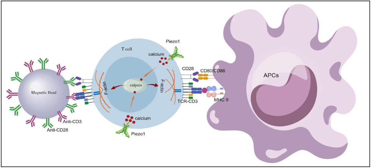
Jiang S, Li H, Zhang L, et al. Generic Diagramming Platform (GDP): a comprehensive database of high-quality biomedical graphics. Nucleic Acids Res. 2025;53(D1):D1670-D1676. doi:10.1093/nar/gkae973
Key words used in this paper:
mechanosensation, Piezo1, T cell receptors(TCR), antigen presenting cells (APCs), T cell activation, immunological synapses.
Things to know before read the paper:
What is mechanosensation/mechanotransduction?
Mechanosensation is a cell’s ability to sense physical forces, such as stretch, pressure, stiffness or shear stress. Cells convert those mechanical forces into biochemical signals that change cell behavior. This is called mechanotransduction.
What are the known mechanosensors?
Piezo1/ Piezo2, TRP channels (TRPV4, TRPV7), TREK-1, ASICs etc.
What is Piezo1?
Piezo 1 is a mechanosensitive, nonselective cation channel that opens in response to membrane stress and allows Ca2+ influx. Piezo1 regulated T cell activation and migration, macrophage function, metabolic reprogramming and other various functions in immune cells as well as non immune cells.
What is T cell and T cell receptor (TCR)?
T lymphocytes are adaptive immune cells that recognize infected cells or abnormal cells and kill those cells. Two major types of T cells: CD4+ helper T cell and Cd8+ cytotoxic T cells.
What are antigen presenting cells (APCs)?
APCs are immune cells that capture antigens, process them into peptides and present them on MHC molecules to T cells (e.g Dendritic cells, Macrophages, B cells).
What is the major histocompatibility Factor (MHC)?
MHC molecules are cell surface proteins that present antigen peptides to T cells. There are two types of MHC molecules: MHC class I and MHC class II. MHC class I molecules are present on all nucleated cells and recognized by CD8+ T cells whereas MHC class II present on professional APCs and recognized by CD4+ T cells.
How are T cells activated?
T cell activation need three major coordinated signals:
- Antigen recognition: TCR binds to peptide MHC complex on APCs and SIGNAL transduction through TCR-CD3 complex
- Co-stimulation: CD28 on T cell binds CD80/CD86 on APCs
- Cytokines: Directs T cell differentiation and fate.
What are antigens?
Antigens are the molecules that can be recognized by the immune system (e.g proteins, peptides, carbohydrates, lipids etc)
What is immunological synapse?
The immunological synapse is the organized contact zone between a T cell and APC where the central ring contains TCR-pMHC complex and the peripheral ring contains adhesion molecules (integrins).
What’s the difference between allogeneic T cell proliferation and autologous T cell proliferation (in this context)?
Autologous T cell proliferation: moDCs and T cells form the same donor. MHC identical, No proliferation of T cells unless treated ( in this paper moDcs are treated with Tetanus toxoid before coculture with T cells).
Allogenic T cell proliferation: moDCs and T cells from different donors, MHC mismatched, triggers strong activation and proliferation, no added antigen required.
Primary Research question:
What is the role of Piezo1 protein in the regulation of the T cell immune response?
Materials used and their functions:
Anti Cd3/CD28 microbeads (Anti Cd3/CD28 Dynabeads): Anti- CD3/CD28 Dynabeads are microscopic magnetic beads coated with antibodies against CD3 and CD28. It provides simultaneous TCR engagements and co-stimulation, inducing robust and sustained T cell activation.
Yoda 1: A small molecular chemical agonist of Piezo1. It opens the Piezo1 mechanosensitive ion channels without mechanical force, causing Ca2+ influx.
Calpain inhibitor (PD150606): Blocks calpains, calcium dependent cystine protease. This prevents cleavage of cytoskeletal and signaling proteins, helping dissect roles of Ca2+ driven proteolysis in T cell activation.
Cyclosporine A(CsA) : A classic calcineurin phosphatase inhibitor. By blocking calcineurin, it prevents NFAT dephosphorylation and nuclear translocation, thereby suppressing T cells activation and cytokine gene transcription.
GM-CSF and IL4: This cytokine duo steers monocytes towards Monocyte-derived dendritic cells (moDCs). GM-CSF supports survival and proliferation, while IL-4 prevents macrophage differentiation and promotes a dendritic cell phenotype.
Piezo 1 si-RNA: Small interfering RNA designed to knock down Piezo1 expression.
EGFP targeting Si RNA: A control si-RNA that targets EGFP, a protein usually absent in most cells. It controls for off-target effects and transfection- related stress, ensuring that observed phenotypes are not due to siRNA delivery itself.
CellTrace dyes (e.g CellTrace Violet or CellTrace CFSE): used for T cell proliferation assays. When a cell divides, the dye is split equally between daughter cells. With every division, fluorescence intensity drops by ~50%.
Flur0-3 AM: Fluo-3 AM is a cell-permeable fluorescent calcium indicator dye used to measure intracellular Ca²⁺ levels in live cells
EGTA: EGTA stands for Ethylene Glycol Tetraacetic Acid. It is a calcium-selective chelating agent used to eliminate extracellular Ca²⁺ from biological solutions and to study Ca²⁺-dependent cellular processes.
Figure decoded:
Now that we know the players (Piezo1, Ca²⁺, calpain, APC-T cell synapse), let’s decode how each figure builds the story. Readers are requested to follow the original article to correlate with our analysis
Figure 1:
- Piezo1 is expressed in human T cells, where it is distributed across the plasma membrane and in intracellular vesicles (figure 1A).
- Piezo1 plays an important role in early T cell activation: Increase phosphorylation of Zap70 and surface expression of CD69 ( early activation marker) when treated with bead immobilized anti Cd3/Cd28 antibodies (figure 1b-E).
- Role of Piezo1 in T cell proliferation: Surface expression of CD25, a proliferation marker, decrease in Piezo1 knockdown cell (figure 1F and 1G). Thus Piezo1 plays an important role in T cell proliferation.
- T cell priming (in both allogeneic and autologous context): Piezo 1 has a crucial role in T cells antigen priming by APCs which is proved by coculturing T cells with both allogeneic and autogenic moDCs ( figure 1H and I).
Figure 2:
- Piezo1 agonist Yoda1, when treated along with soluble CD3/CD28 (alone less efficient in activating T cell) induced high level of T cell activation which is independent of mechanical stress. It significantly increases the expression of activation marker CD69 (figure 2A)and the phosphorylation of Zap70 (figure 2B).
Figure 3:
- Piezo1 is distributed not centrally but peripherally in the immunological synapse (figure 3A and B).
- Upon TCR activation ( by both bead bound and soluble CD3/CD28) intracellular Ca2+ level increased in control T cells in comparison with Piezo1 knockdown T cells (figure 3C).
- Addition of Yoda1 with soluble CD3/CD28, also increases the Ca2+ level (figure D). Further addition of EGTA, an extracellular Ca2+ chelating agent, decreases Yoda1 induced intracellular Ca2+ level (figure E and F).
Figure 4:
- Addition of calpain inhibitor in presence of Yoda1 reduced the expression of CD69,thus abolishing optimal TCR activation (figure 4A). Whereas addition of calcineurin phosphatase did not decrease CD69 expression (figure 4B). This Piezo1 dependent T cell activation is primarily calpain dependent.
- Furthermore F- actin increased during T cell activation in presence of Yoda1 which is decreased in addition of calpain inhibitor
- The confocal microscopy image showed the colocation of Piezo1 with F actin because of its peripheral distribution in immunological synapses.
Take home massage from this paper:
Piezo1 is a specialized mechanosensors on human T cells that is essential for translating mechanical forces into optimal immune activation. Piezo1 acts as a physical-to-chemical bridge, ensuring that the T cell only “turns on” fully when it experiences the correct mechanical interaction with another cell.
Limitations:
- No in vivo validation: The study lacks animal models or clinical data demonstrating that Piezo1 regulates T cell activation during real immune responses such as infection, vaccination, or autoimmunity
- Mechanical forces not directly quantified : Mechanical stress at the immunological synapse is inferred rather than directly measured. There is no direct quantification of force magnitude, membrane tension, or biophysical parameters activating Piezo1.
- Partial Piezo1 knockdown: Piezo1 was reduced using siRNA, achieving roughly 40–60% knockdown. Residual Piezo1 activity may mask stronger phenotypes, and complete genetic deletion effects remain unknown.
Future Direction:
- Quantitative mechanobiology of the immunological synapse: Future studies should directly measure mechanical forces at the TCR–pMHC interface using tools such as traction force microscopy, optical tweezers, or molecular tension sensors to define the force thresholds required for Piezo1 activation.
- Cross-talk with other mechanosensors and cytoskeletal regulators: Future work should assess whether Piezo1 cooperates with integrins, actomyosin tension, or other mechanosensitive ion channels to orchestrate force-dependent immune signaling.
Role of tissue stiffness and microenvironment mechanics
Investigating how extracellular matrix stiffness and tissue mechanics influence Piezo1 activation may uncover how physical niches tune immune responses in lymph nodes, tumors, and inflamed tissues.

Editor, Research Paper Decoded