
What if a tiny change in DNA could influence how lung cancer is treated?
Imagine searching for one person in a stadium filled with thousands of people. You know exactly who you are looking for. But everyone around them looks almost identical. Now imagine that the person you are searching for represents a tiny change in DNA. That is one of the challenges scientists face when trying to detect genetic mutations associated with cancer.
DNA carries the instructions that help our cells function. Sometimes, a small change in those instructions can contribute to the development of cancer or influence how a cancer responds to treatment. But finding that change is not always straightforward. A blood sample from someone with cancer may contain thousands of DNA fragments from normal cells, while only a small fraction comes from the tumor. The question is:
How do you find a rare cancer-associated mutation when normal DNA is everywhere?
In a 2026 study published in Analytical Chemistry, researchers explored a strategy combining DNA amplification, a molecular blocking technique and a protein-based detection system to identify specific mutations associated with non-small cell lung cancer.
Their approach brings together three molecular tools:
- LAMP, which amplifies DNA.
- A blocker primer, designed to suppress amplification of normal DNA.
- PfAgo, a protein used to recognize target DNA and generate a detectable signal.
Let’s understand why this approach matters—and how it works.
1. First, why are scientists interested in EGFR mutations?
To understand the research, we need to start with the disease.
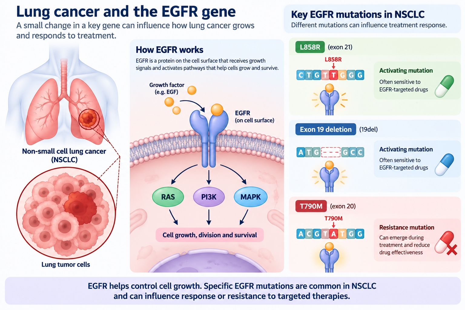
Lung cancer is not a single disease. It includes different types of tumors that can behave differently and respond differently to treatment. One major category is non-small cell lung cancer (NSCLC), which accounts for approximately 85% of lung cancer cases. Within NSCLC, scientists have identified genetic changes that can help explain how cancer cells grow and respond to particular medicines. One important gene is called EGFR.
What is EGFR?
EGFR stands for epidermal growth factor receptor. Think of it as a molecular switch involved in cell growth and survival. Under normal conditions, EGFR receives signals that help cells grow, divide and respond to their surroundings. But certain mutations can alter how this signaling system works.
Some EGFR mutations can keep growth-related signaling active, helping cancer cells survive and multiply. This is why EGFR mutations are important in lung cancer: identifying the specific mutation can provide information that helps clinicians select targeted treatment.
Three mutations, three important stories
The researchers focused on three EGFR alterations:
EGFR L858R
This is a point mutation in exon 21. A single amino-acid change alters the EGFR protein and can make tumors sensitive to certain EGFR-targeted medicines.
EGFR exon 19 deletion (19del)
Here, a small section of DNA is deleted from exon 19. These deletions are also associated with sensitivity to particular EGFR inhibitors.
EGFR T790M
This mutation is especially important in the treatment-resistance story. Some patients initially respond to earlier-generation EGFR-targeted medicines. Over time, cancer cells may acquire changes that allow them to escape treatment. T790M is a well-known resistance-associated mutation that can emerge during treatment with earlier-generation EGFR inhibitors. Detecting it can help inform treatment decisions, including consideration of medicines such as Osimertinib.
The important point is that the mutation is not simply a genetic label. It can provide information about the biology of the tumor and its relationship with treatment.
2. The problem: cancer DNA can be hidden among normal DNA
Now that we understand why EGFR mutations matter, let’s look at the diagnostic challenge. Traditionally, doctors may obtain tumor tissue through a biopsy to investigate the cancer and identify relevant genetic alterations. A biopsy can provide valuable information, but obtaining tissue may be invasive. It may also be difficult to repeat frequently, especially when doctors want to monitor changes over time. This is where liquid biopsy becomes interesting.
What is a liquid biopsy?
Cancer cells can release small fragments of DNA into the bloodstream. These fragments are called circulating tumor DNA (ctDNA). A blood sample can therefore contain:
- DNA released by normal cells.
- DNA released by cancer cells.
- A mixture of genetic material from different sources.
This means scientists may be able to investigate tumor-associated mutations using a blood sample rather than relying only on tumor tissue. But there is a catch.
The amount of tumor-derived DNA can be very small. In some situations, ctDNA may represent less than 1% of the total cell-free DNA in plasma. The proportion can be especially low in early-stage disease or when only a small amount of tumor DNA is being released. Imagine a sample containing 100,000 DNA fragments. If only a small number carry the mutation you are looking for, detecting them becomes difficult. And if the normal DNA looks almost identical to the mutated DNA, the challenge becomes even greater.
The problem is not simply finding DNA. It is finding the right DNA among a much larger background.
3. Why ordinary DNA amplification may not be enough
Scientists often use a technique called DNA amplification. The idea is simple. If a sample contains only a small amount of DNA, researchers can make many copies of a specific genetic sequence. This is similar to photocopying a page from a book. Instead of working with one copy, you create many copies so that the information becomes easier to detect. One widely used amplification technique is PCR. Another is LAMP.
What is LAMP?
LAMP stands for loop-mediated isothermal amplification. Unlike conventional PCR, which repeatedly changes temperature to separate and copy DNA, LAMP works at a relatively constant temperature.
This makes it attractive for diagnostic systems that aim to reduce dependence on complex thermal cycling equipment. But there is a problem when the target is a rare mutation. Suppose a sample contains:
- A large amount of normal EGFR DNA.
- A very small amount of mutated EGFR DNA.
If the amplification reaction copies both sequences, the normal DNA may dominate the reaction. The mutation is still present, but its signal can become difficult to distinguish from the background. The researchers observed this challenge when conventional LAMP produced amplification from wild-type DNA under their experimental conditions.
So they needed something more selective. Not just a method that makes more DNA. A method that helps the mutated DNA stand out.
4. The clever idea: block the normal DNA, amplify the mutant
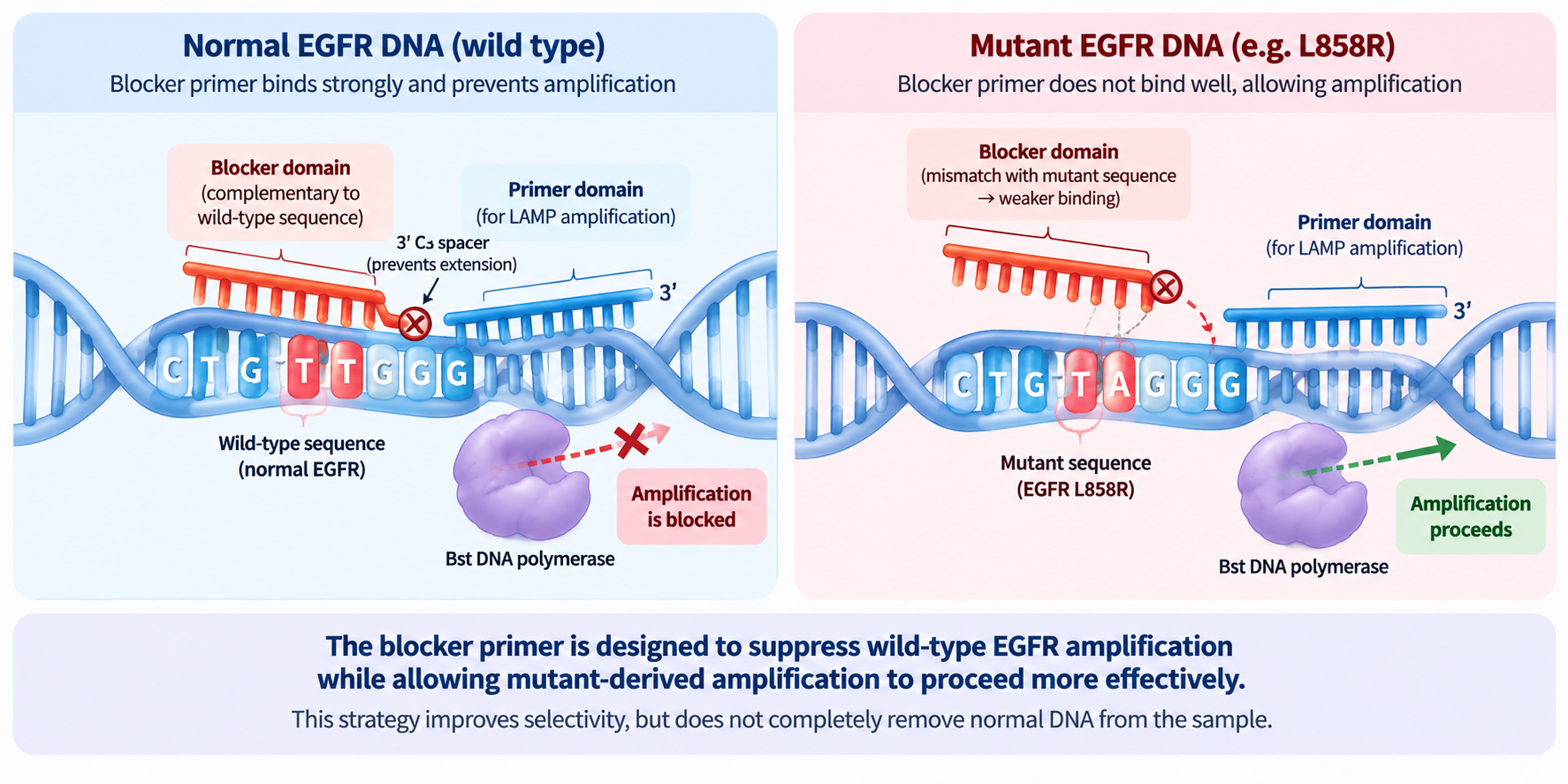
This is where the blocker primer enters the story. A blocker primer is a DNA-based tool designed to interfere with amplification of a particular sequence. In this study, the researchers designed a blocker primer that binds strongly to the normal, or wild-type, EGFR sequence. The goal was to suppress amplification of the normal DNA.
But what happens when the DNA contains the mutation? The sequence is no longer a perfect match for the blocker. That difference can reduce blocker binding and allow amplification of the mutant target to proceed. In simple terms:
Normal DNA → blocker binds → amplification is suppressed
Mutant DNA → blocker binding is disrupted → amplification can proceed
This strategy is intended to give the rare mutant DNA a better chance of being detected. It does not mean that normal DNA disappears from the sample. Instead, the reaction is designed to reduce the contribution of normal DNA during amplification. That distinction is important. The researchers are not physically removing all the normal DNA. They are trying to make the amplification process more selective.
Why this matters
If a mutation is present at a very low frequency, even a small improvement in selectivity can make a difference. The blocker primer helps address the first part of the problem:
How can we reduce the overwhelming contribution of normal DNA?
But the researchers still need a way to recognize the amplified mutation and convert it into a measurable signal. That brings us to the next molecular tool.
5. Meet PfAgo: the molecular investigator
After amplification, the researchers used a protein called PfAgo. Its full name is Pyrococcus furiosus Argonaute.
That sounds complicated, but its role in this study can be understood through a simple analogy. Imagine that DNA amplification has produced many copies of the target sequence. Now you need a molecular investigator that can recognize the sequence you are interested in. PfAgo is used for this recognition and cleavage step.
How does PfAgo recognize the target?
The researchers used short DNA guides. These guides help direct PfAgo toward the target sequence. When the appropriate target is recognized, PfAgo can cleave the DNA at a specific site. The cleavage event is important because it helps generate DNA fragments that participate in the next stage of the detection process. The researchers designed the system so that these fragments could act as additional guides. This creates a cascade.
One recognition event can help generate more guide molecules, which can activate additional PfAgo molecules. The system then uses fluorescent reporter molecules. When the reporter molecules are cleaved, the fluorescence signal changes. That signal can be measured. So the detection process has two important layers:
- Recognition: PfAgo identifies and cleaves the target-associated DNA.
- Signal generation: The cleavage cascade activates fluorescent reporters, producing a measurable signal.
The goal is to make a small amount of target DNA easier to detect.
6. Putting the molecular pieces together
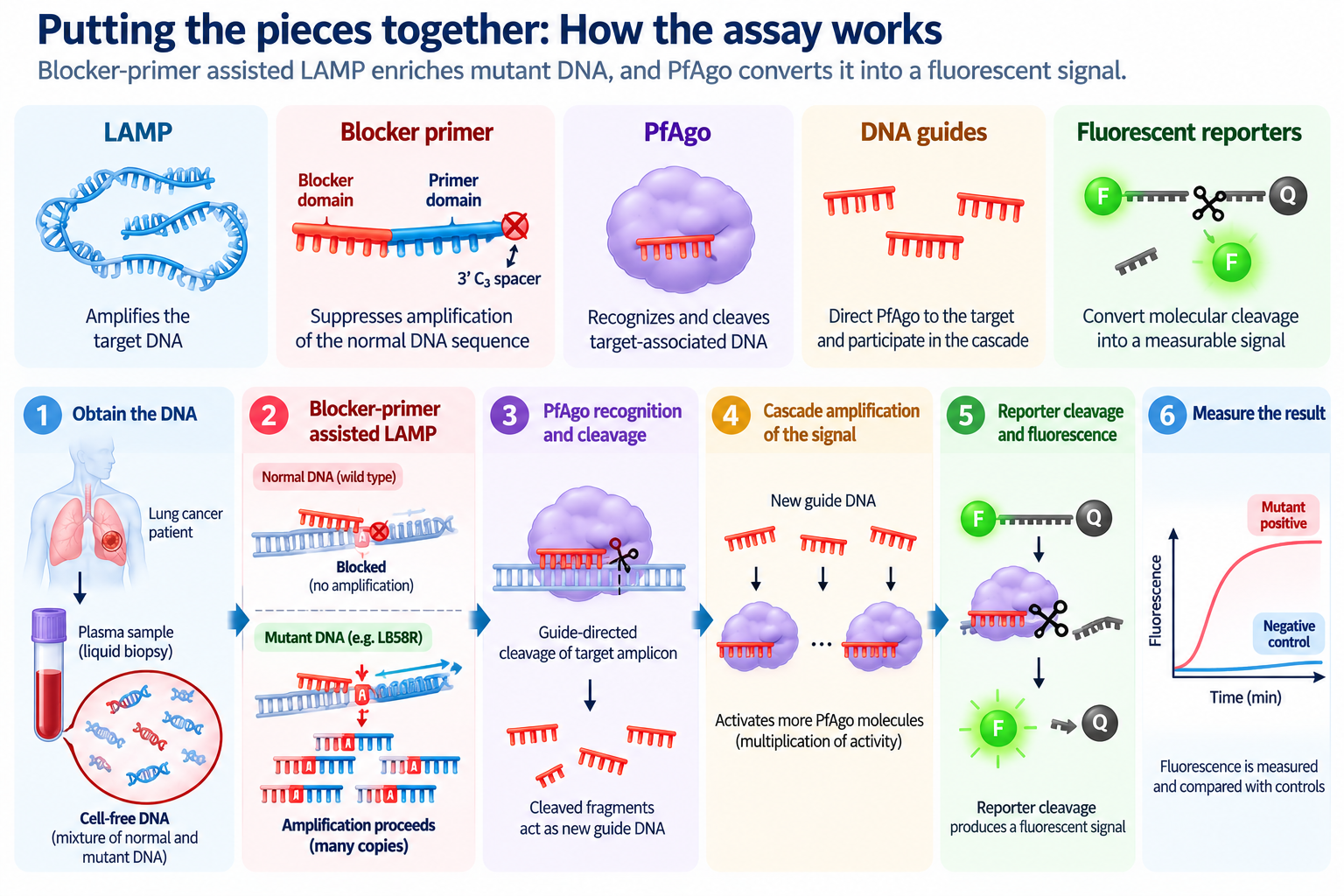
Now let’s connect the entire strategy. The researchers combined the blocker-primer approach with LAMP and PfAgo. Each component has a different job.
| Component | What it does |
|---|---|
| LAMP | Amplifies the target DNA |
| Blocker primer | Suppresses amplification of the normal DNA sequence |
| PfAgo | Recognizes and cleaves target-associated DNA |
| DNA guides | Direct PfAgo toward the target sequence and participate in the cascade |
| Fluorescent reporters | Convert molecular cleavage into a measurable signal |
The workflow can be understood as a sequence of events.
Step 1: Obtain the DNA
The researchers worked with DNA from lung cancer cell lines and cell-free DNA extracted from plasma samples.
Step 2: Amplify the target
LAMP produces many copies of the target sequence.
Step 3: Suppress normal DNA amplification
The blocker primer is designed to reduce amplification of the wild-type sequence.
Step 4: Recognize the mutation
PfAgo, directed by DNA guides, recognizes and cleaves the target-associated DNA.
Step 5: Amplify the signal
The cleavage products help trigger additional guide-mediated activity.
Step 6: Measure fluorescence
The fluorescent reporter molecules generate a signal that can be measured. The researchers then compare the signal with controls to determine whether the target mutation is detected.
7. Could the method detect a mutation when very little mutant DNA was present?
This is where the study becomes particularly interesting. The researchers evaluated the assay using mixtures containing mutant DNA and a large background of wild-type DNA. The total DNA concentration in the sensitivity experiments was 1 × 10⁶ copies/µL. They tested different mutant allele frequencies.
What is mutant allele frequency?
Mutant allele frequency, or MAF, describes the proportion of DNA copies carrying the mutation within the measured DNA population. For example:
If 1 out of 100 DNA copies carries the mutation, the mutant allele frequency is 1%.
If 1 out of 1,000 copies carries the mutation, the frequency is 0.1%.
A lower frequency means the mutation is harder to detect because it is surrounded by more normal DNA.
The reported analytical sensitivity
The researchers reported detection of:
- EGFR L858R
- EGFR T790M
- EGFR exon 19 deletion
at mutant allele frequencies as low as 0.05% in their experimental high-wild-type background.
That means the assay was able to detect a very small mutant fraction under the study’s analytical conditions. The researchers also compared the method with conventional LAMP. Conventional LAMP produced evident nonspecific amplification from wild-type DNA under the conditions tested. The blocker-primer-assisted strategy reduced this background and improved mutation discrimination. This is the central analytical finding of the study. However, there is an important distinction:
Analytical sensitivity is not the same as clinical sensitivity.
Detecting a mutation in a carefully prepared DNA mixture does not automatically mean the same detection limit will be achieved in every patient’s blood sample. Clinical samples introduce additional challenges, including variable tumor DNA abundance and differences in DNA recovery.
8. What happened when the researchers tested lung cancer cell lines?
The researchers also tested DNA from three lung cancer cell lines with known EGFR mutation profiles. These were:
A549: Used as a wild-type EGFR control for the mutations examined.
HCC827: A cell line carrying an EGFR exon 19 deletion.
H1975: A cell line carrying EGFR L858R and T790M.
The assay produced stronger signals for the expected mutations in H1975 than in the wild-type and alternative-mutation controls. This supported the idea that the method could distinguish the target mutations in genomic DNA from different lung cancer cell lines. Why is this useful?
Because purified DNA mixtures are not the only way to evaluate a molecular diagnostic method. Cell-line genomic DNA provides a more complex genetic background and helps test whether the assay can recognize its target among other DNA sequences. But cell lines are still laboratory models. They do not fully reproduce the biological variability of patient samples. That is why the researchers also investigated clinical plasma.
9. The clinical question: could it detect EGFR L858R in patient plasma?
The researchers evaluated the assay using cell-free DNA extracted from plasma samples from patients with NSCLC. The clinical comparison focused on EGFR L858R. They tested 25 plasma samples:
- 13 samples confirmed positive for L858R by next-generation sequencing.
- 12 samples confirmed negative by next-generation sequencing.
Next-generation sequencing, or NGS, was used as the reference method. NGS can examine many DNA sequences and identify genetic alterations in a sample. The researchers compared the results from their assay with the NGS results.
What did they find?
| Measurement | Study result |
|---|---|
| NGS-positive samples | 13 |
| NGS-negative samples | 12 |
| True positives detected | 11 |
| False negatives | 2 |
| True negatives | 12 |
| False positives | 0 |
| Sensitivity | 84.62% |
| Specificity | 100% |
| AUC | 0.9231 |
The assay detected 11 of the 13 NGS-positive samples. Two positive samples were not detected. All 12 NGS-negative samples were classified as negative by the assay. The researchers suggested that the false-negative results may have been related to very low mutant abundance in the plasma DNA or variability in DNA recovery.
What do these numbers mean?
Sensitivity describes how often the assay correctly identifies samples that are positive according to the reference method. In this study, the assay detected 84.62% of the NGS-positive samples.
Specificity describes how often the assay correctly identifies samples that are negative according to the reference method. The study reported 100% specificity in this 12-sample negative group.
That is encouraging, but the sample size was small. The result should not be interpreted as proof that the assay will have 100% specificity in a larger or more diverse patient population. The study provides preliminary clinical evidence—not a completed clinical validation.
10. Why is this approach interesting for molecular diagnosis?
The research brings together three ideas.
First: amplification is not always enough
Making more copies of DNA does not automatically solve the problem of identifying a rare mutation. If normal DNA is amplified along with the mutant DNA, the target can remain difficult to distinguish.
Second: selectivity matters
The blocker primer is designed to reduce wild-type amplification. This helps address the imbalance between abundant normal DNA and rare mutant DNA.
Third: signal amplification can help
PfAgo provides a target-recognition and cleavage step that is connected to a fluorescent signal cascade. The combination allows the researchers to address both the amplification problem and the signal-detection problem.
That is what makes the strategy interesting. It is not simply another DNA amplification reaction. It is an attempt to combine selective amplification with molecular signal amplification.
The authors also describe the method as operating under isothermal conditions, which may be useful for developing diagnostic systems with less dependence on conventional PCR thermal cycling. But the study does not establish that the method is already a rapid, inexpensive, point-of-care lung cancer test. That would require further work.
11. What are the limitations?
Every diagnostic method has limitations, and this study is no exception.
The clinical sample size was small
The clinical plasma evaluation included 25 samples. A larger study would be needed to establish performance across a wider range of patients and mutation abundances.
Two positive samples were missed
The assay did not detect two samples that were positive for L858R by NGS. This matters because a negative result from a molecular test does not necessarily mean that the mutation is absent.
The analytical detection limit does not equal clinical detection performance
The reported 0.05% mutant allele frequency was obtained under controlled analytical conditions. It should not be interpreted as a guaranteed detection limit for every plasma sample.
The clinical evaluation focused on one mutation
Although the assay was investigated for L858R, T790M and exon 19 deletion, the clinical plasma comparison reported in the paper focused on L858R. Further clinical evaluation would be required for the other mutations.
It is not a replacement for established clinical testing
The study demonstrates a promising experimental approach. It does not establish that the assay can replace NGS or other validated clinical tests for treatment decisions. More extensive validation, including larger patient cohorts and assessment of clinical utility, would be necessary.
12. The bigger picture: detecting the mutation is only the beginning
The most interesting part of this research is not simply that the assay detected a mutation at a low allele frequency. It is the strategy behind it.
The researchers recognized that rare mutation detection has two connected challenges:
- The target DNA may be overwhelmed by normal DNA.
- Even after amplification, the target needs to be recognized and converted into a measurable signal.
Their approach addresses these challenges through a combination of blocker-primer-assisted LAMP and PfAgo-mediated signal amplification. The results suggest that this combination can detect selected EGFR mutations in controlled experiments and can identify L858R in a preliminary plasma sample cohort. That makes it an interesting direction for further research into liquid biopsy-based mutation detection.
But the next question is just as important: Can the method maintain its analytical performance across larger, more diverse clinical samples—and can it provide reliable results that meaningfully support patient care?
That is what future studies will need to establish.
Take-home message
A rare cancer-associated mutation can be difficult to detect when it is surrounded by a much larger amount of normal DNA.
This study explored a strategy that combines LAMP amplification, a blocker primer designed to suppress wild-type DNA amplification, and PfAgo-mediated recognition and fluorescent signal amplification.
The researchers reported analytical detection of EGFR L858R, T790M and exon 19 deletion at mutant allele frequencies as low as 0.05% under their experimental conditions.
In a preliminary plasma comparison, the assay detected 11 of 13 NGS-positive L858R samples and correctly classified all 12 NGS-negative samples. The findings are promising, but larger clinical studies are needed before the approach can be considered for routine diagnostic use.
The lesson is simple: in molecular diagnosis, making more DNA is not always enough. Sometimes the real challenge is making the right DNA stand out.
PaperDecoded — Making molecular research easier to understand, one paper at a time.
Original Research paper
Yuan J, Su G, Weng B, et al. Blocker Primer-Assisted LAMP Coupled with Pyrococcus furiosus Argonaute Enables Specific and Sensitive Detection of EGFR Mutations in NSCLC. Analytical Chemistry. 2026;98:24357–24365.
DOI: https://doi.org/10.1021/acs.analchem.6c03086

Editor, Research Paper Decoded